Browser update
You're using an old browser, please update your browser. For more information visit
Optimization and compatibility
.
EN
PT
ES
About
Atlas
Videos
News
Search...
Home
Atlas
OPTIC NERVE DISEASES
Optic Disc Pits and Associated Serous Macular Detachments
Clinical Case 01
Search
Free text search
Diseases
Clinical Cases
Chapters
all chapters
AGE-RELATED MACULAR DEGENERATION
CHORIORETINAL INFLAMMATORY DISEASES - Infectious Causes
CHORIORETINAL INFLAMMATORY DISEASES - Noninfectious Causes
CONGENITAL AND PEDIATRIC RETINAL DISEASES
DIABETIC RETINOPATHY
MACULAR DISEASES
OPTIC NERVE DISEASES
PERIPHERAL RETINAL DISEASES
RETINAL AND CHOROIDAL TUMORS
RETINAL DYSTROPHIES AND SYNDROMES
RETINAL VASCULAR DISEASES
TRAUMATIC RETINOPATHIES
UNUSUAL CLINICAL CASES
VITREOUS DISEASES
Clinical Cases
Age range
-
Instants range
-
Exam type
All exam types
Angiography with Green Indocyanine
Anterior Segment Photo
Autofluorescence
Biometry
Colour Retinography
Computed Tomography
Contrast sensitivity / Colour sensitivity
Corneal Topography
Echography
ERG, multifocal
ERG, pattern
Fluorescein Angiography
Infrared
Microperimetry
Nervous Fiber Analysis
OCT B-Scan (Longitudinal)
OCT C-Scan (Coronal)
OCT, Cornea
Perimetry
Red-free
RNFL - Nervous Fiber Analysis
RNFL - Thickness Image
Specular Microscopy
Surgical Video
Tonometry
has images
has videos
New
OPTIC NERVE DISEASES
Optic Disc Pits and Associated Serous Macular Detachments
Clinical Case 01
Next
Start
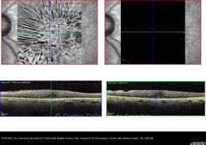
OCT horizontal line-scan shows a neurosensorial detachments and a bilaminar structure "schisis-like" between external and internal retinal layers associated to an optic disc pit
End
OCT horizontal line-scan showing total recovery of retinal structure after a new surgical technique. Visual acuity: 20/20 LE
Instants
162
2
Display #
4
8
12
20
Order results
Default order
# Exams
# Images
# Videos
Instant date
Created date
Ascending
Descending
1
2
Results 1-8 of 11
1st evaluation
5
10
0
Instant Images
04-02-2010
Colour Retinography
3
23-02-2010
Red-free
1
23-02-2010
Autofluorescence
1
23-02-2010
Infrared
1
23-02-2010
OCT B-Scan (Longitudinal)
4
2nd evaluation - postoperative
3
13
1
Instant Images
24-02-2010
Surgical Video
16-03-2010
Anterior Segment Photo
1
16-03-2010
Colour Retinography
1
16-03-2010
OCT B-Scan (Longitudinal)
11
3rd evaluation - postoperative
5
18
1
Instant Images
18-03-2010
Surgical Video
19-03-2010
Colour Retinography
1
12-04-2010
Red-free
1
12-04-2010
Autofluorescence
1
12-04-2010
Infrared
1
12-04-2010
OCT B-Scan (Longitudinal)
14
4th evaluation
5
18
0
Instant Images
19-04-2010
OCT B-Scan (Longitudinal)
11
26-04-2010
Colour Retinography
1
26-04-2010
Red-free
2
26-04-2010
Autofluorescence
2
26-04-2010
Infrared
2
5th evaluation
4
21
0
Instant Images
15-02-2011
Colour Retinography
2
15-02-2011
Red-free
2
15-02-2011
Autofluorescence
2
15-02-2011
OCT B-Scan (Longitudinal)
15
6th evaluation
5
17
0
Instant Images
17-05-2011
Colour Retinography
2
17-05-2011
Red-free
1
17-05-2011
Autofluorescence
1
17-05-2011
Infrared
1
17-05-2011
OCT B-Scan (Longitudinal)
12
7th evaluation
6
28
0
Instant Images
01-01-1970
Red-free
1
22-05-2012
Anterior Segment Photo
1
22-05-2012
Colour Retinography
3
22-05-2012
Autofluorescence
1
22-05-2012
Infrared
1
22-05-2012
OCT B-Scan (Longitudinal)
21
8th evaluation
5
12
0
Instant Images
22-01-2013
Autofluorescence
2
22-01-2013
Red-free
1
22-01-2013
Infrared
1
22-05-2013
Colour Retinography
2
22-05-2013
OCT B-Scan (Longitudinal)
6
Results 1-8 of 11
1
2
Next