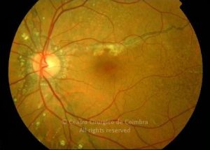
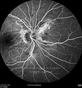
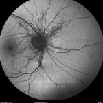
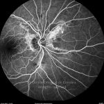
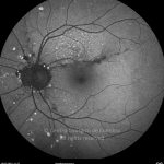
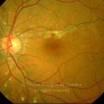
Angioid streaks are usually bilateral and represent visible breaks in the Bruch’s membrane. The elastic fibers of Bruch’s membrane are charged with calcium, and this accumulation makes the layer brittle and susceptible to breaks. Clinically they consist of dark red to brown linear bands of irregular contour, originating radially from the optic disc and extending into the periphery. Near the optic disc Retinal pigment epithelium contiguous to the borders of the streaks, can be hypopigmented or thinned The transition regions between normal and pathological retina may have a fine stippled appearance, known as “peau d’orange”. In the mid-periphery, small depigmented subretinal deposits, known as crystalline spots, are frequently present. Other ocular findings include optic disc drusen and peripheral round atrophic scars.
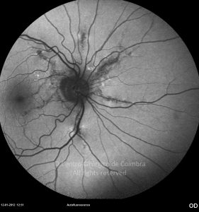
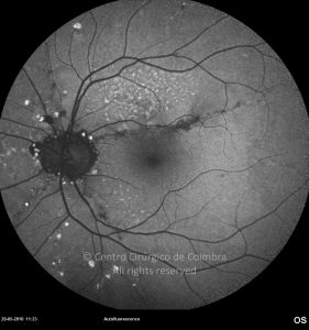
In FAF, recent angioid streaks produce no changes in the autofluorescence pattern. Chronically, as the RPE/photoreceptor complex becomes atrophic, irregular hypofluorescent lines with an enhanced autofluorescence border appear.
The most frequent association with systemic disease is pseudoxanthoma elasticum (PXE) also known as Gronblad-Strandberg syndrome. Several other systemic diseases have been associated with angioid streaks (see below) however similar changes can be seen in healthy older patients.
Patients are usually asymptomatic. Angioid streaks can, however cause subretinal hemorrhages associated or not with CNV. When CNV is present, therapeutic options include Laser (if the lesion is extrafoveal), PDT or intravitreal injection of an anti-VEGF agent.


