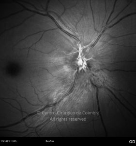
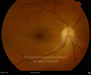
Anterior ischemic optic neuropathy is an ischemic damage of the optic nerve head. It is characterized by a painless unilateral visual loss, developing over a period of hours to days. Relative afferent pupillary defect is usually present, unless there is bilateral optic neuropathy. A visual field defect is usually found, most commonly altitudinal or arcuate.
An important distinction must be made between two clinical pictures that have different therapeutic and prognostic implication: arteritic anterior ischemic optic neuropathy (AAION) and non-arteritic anterior ischemic optic neuropathy (NAION).
AAION is the less frequent of the two entities, corresponding to 5-10% of the cases of AION. It is a serious condition corresponding to the occlusion of the short posterior cilliary arteries by an inflammatory process. The mean age of diagnosis is 70 years, or 10 years more than NAION.
A good clinical history is fundamental in this group of patients. NAION is frequently accompanied by malaise, anorexia and weight loss, fever, joint and muscle pain. Headache and scalp tenderness in the temporal artery territory may be present. The most specific symptom is jaw claudication. A commonly associated syndrome is that of polymyalgia rheumatic. Visual acuity is usually severely compromised, being< 20/200 in 60% of patients.
Fundus examination usually reveals a pale optic disc edema. Cotton wool spots may be present. Choroidal ischemia is frequent and manifests as peripapillary pallor and edema and delayed choroidal perfusion in fluorescein angiography. Erythrocyte sedimentation rate, C-reactive protein, leukocyte count and alkaline phosphatase levels are usually elevated, while erythrocyte count and hemoglobin levels are usually depressed.
Diagnosis is confirmed with temporal artery biopsy, showing inflammatory destruction of media-intima junction, with or without giant cells. A positive biopsy is diagnostic; however a negative one does not exclude the diagnosis.
Once there is a high clinical suspicion, treatment should be initiated promptly with a high dose corticosteroid.
Prompt treatment is intended not to improve the vision of the affected eye, but to avoid systemic vascular complications as well as to preserve the fellow eye (involved in up to 95% of patients in the following days to weeks if untreated).
NAION is more common, corresponding to 90-95% of the cases of AION. The mean age of diagnosis is 60 years. Visual loss is usually less severe than that of AAION with >60% of patients having visual acuities greater than 20/200. Patients usually report visual loss noted on awakening. Fundus examination reveals an optic disc edema, diffuse or sectorial, pale or hyperemic. The observation of the fellow eye usually reveals a small optic disc, with a small or absent physiologic cup. Usually the optic disk becomes atrophic after 4-8 weeks. Fluorescein angiography demonstrates a delayed optic disc filling in 75% of the cases. Crowding of the optic disc, hypertension, diabetes, smoking and hyperlipidemia are demonstrated risk factors for NAION.
There is no proven treatment for NAION or for prophylaxis of the fellow eye.


