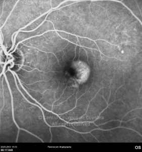
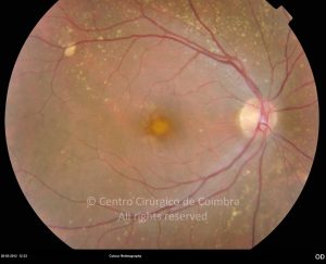
Best vitelliform macular dystrophy (VMD), or Best disease, is an autosomal-dominant disorder with variable penetrance and expressivity, characterized by variable deposition of yellowish material (lipofuscin) in the retinal pigment epithelium (RPE) and/or subretinal space.
Onset may occur in childhood or decades later. In the “previtelliform” stage, there is a normal fundus appearance. The electro-oculogram (EOG) is usually abnormal, even in asymptomatic patients and is therefore helpful in making the diagnosis.
The disease is slowly progressive and eventually results in atrophy of the retinal pigment epithelium (RPE) and photoreceptors, severely impairing central vision. Fundus autofluorescence is useful in detecting the lipofuscin accumulation which appears hyperautofluorescent. Patients may complain of blurred vision, poor visual acuity, or metamorphopsia and the symptoms may worsen if the disease progresses to the atrophic stage. Choroidal neovascularization and hemorrhagic detachment of the macula may occur at each stage.
Mutations in the VMD2 gene coding for bestrophin, mapped to 11q12-q13.1, have been associated with Best disease. To date, there is no treatment for this disorder.
Differential diagnosis: adult vitelliform macular dystrophy (pattern dystrophy), basal laminar drusen, acute exudative vitelliform maculopathy, Stargardt disease with large central flecks and idiopathic macular telangiectasias.


