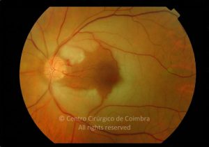
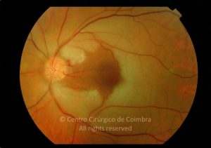
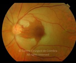
 Fundus photograph of a central retinal artery occlusion occuring 8 days before this evaluation in a 60-years-old female patient. Note the cilioretinal artery maintains a small area of normal appearing retina between the disc and the fovea. The remaining retina has a typical whitish appearance. Visual acuity: 20/200 LE


 Fundus photograph of a central retinal artery occlusion occuring 8 days before this evaluation in a 60-years-old female patient. Note the cilioretinal artery maintains a small area of normal appearing retina between the disc and the fovea. The remaining retina has a typical whitish appearance. Visual acuity: 20/200 LE
