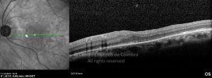
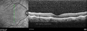
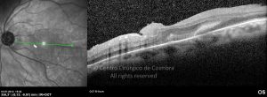
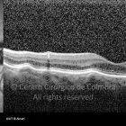
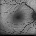
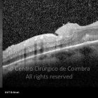
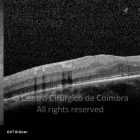
Chorioretinal folds results from compressive forces on the sclera that produce a series of folds in the inner choroid, Bruch´s membrane, RPE and retina. They may be idiopathic or may be secondary to tumors, hypotony, inflammation (posterior scleritis, idiopathic orbital inflammation, and autoimmune disorders), choroidal neovascular membranes, papilledema, and scleral buckle.
The patients are usually asymptomatic or may have metamorphopsia and decreased vision if folds involve fovea.
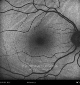
The folds appear as alternating bright peaks and dark troughs or valleys predominantly involving the posterior pole. They may be curvilinear, parallel, or circular. Idiopathic folds are usually bilateral and symmetric. Fundus autofluorescence may be useful in detecting the presence of such folds that result from stretching and thinning of the RPE. Similarly, the fluorescein angiogram shows alternate bands of hyper and hypofluorescence.
The most common cause of choroidal folds is disciform scarring of the macula from neovascular age-related macular degeneration.
Differential Diagnosis:
- Retinal folds due to an epiretinal membrane
- Rhegmatogenous retinal detachment
- Toxocariasis
- Retinopathy of prematurity


