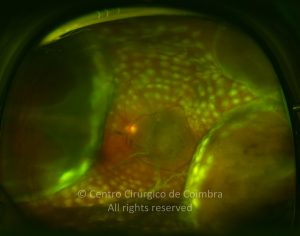
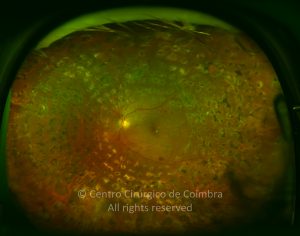
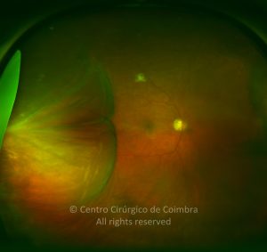
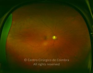
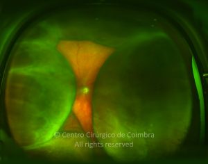
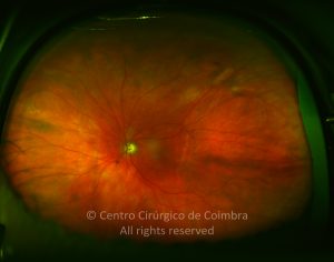
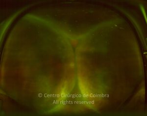
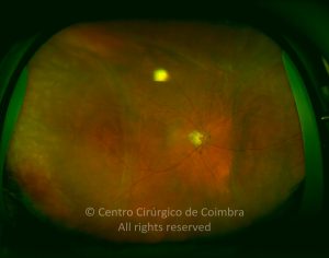
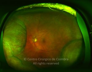
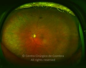
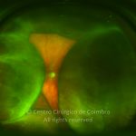
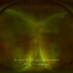
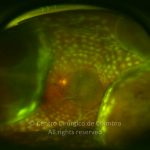
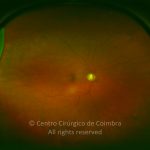
The choroidal detachment presents as a smooth, bullous, orange-brown elevation of the retina and choroid, usually extending 360 degrees around the periphery in a lobular configuration. The ora serrata is visible without scleral depression.
The suprachoroidal space is normally virtual because the choroid is in close apposition to the sclera. As fluid accumulates, this space becomes real, and the choroid is displaced from its normal position. Fluid accumulation, either a serum-like liquid or blood, can also occur within the choroid, which is a spongy tissue.
As a sequela, linear areas of pigmented epithelium hypertrophy, called Verhoeff lines, indicate the posterior limits of the choroidal detachment after fluid reabsorption.
Choroidal detachment may occur in two forms: Choroidal Effusion: Serous choroidal detachment involves transudation of serum into the suprachoroidal space; it is associated with acute ocular hypotony, post-surgical situations, posterior scleritis, Vogt-Koyanagi-Harada syndrome, trauma, intraocular tumors, or uveal effusion syndrome. Choroidal Hemorrhage: Rupture of choroidal vessels causes bleeding into the suprachoroidal space. It can arise spontaneously (rare), as a consequence of ocular trauma, during or after eye surgery.


