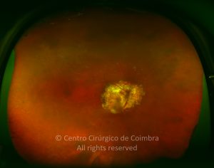
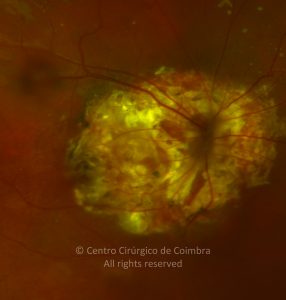
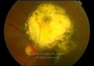
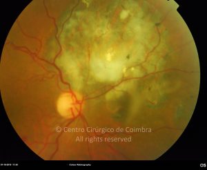
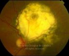
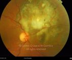
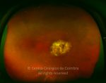
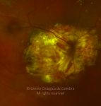
Choroidal hemangioma is a vascular hamartoma and can exist in two distinct forms: the diffuse and the circumscribed type, which differ clinically and histopathologically.
The circumscribed choroidal hemangioma is seen as a well defined red-orange, slightly elevated dome-shaped lesion usually located at the posterior pole.
It usually exhibits slow enlargement in early life, and by young adulthood overlying atrophic changes in the RPE can develop, as can cystoid degeneration of the sensory retina, and sensory retinal detachment.
The diffuse choroidal hemangioma, which permeates throughout the entire choroid, is frequently associated with facial hemangioma (Sturge-Weber disease).


