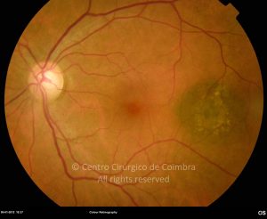
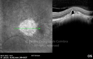
Choroidal nevus is a benign melanocytic tumour, which presents itself as a flat, dark gray-brownish pigmented lesion, occurring more commonly in the post-equatorial region, often with overlying drusen and a yellowish ring around the base (halo nevus).
Multiple nevi may occur in patients with neurofibromatosis.
The nevus can grow during puberty, but in adults the growth should be watched. The chance of a nevus converting into a melanoma is extremely low (1/20,000). Features such as subretinal fluid or overlying orange pigment imply an active mass and could be an early sign of a malignant transformation of the lesion.
Choroidal neovascularization membrane associated with exudation or hemorrhage may develop.
The features in the fluorescein angiogram will vary depending on the degree of pigmentation. When the nevi encroaches or replaces part of the choriocapillaris, the lesions may appear hypofluorescent.
Differential Diagnosis:
- Choroidal melanoma
- Combined hamartoma
- Congenital hypertrophy of the RPE
- Hyperplasia of the RPE
- Subretinal hemorrhage.


