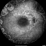
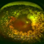
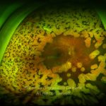
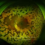
Gyrate atrophy is a generalized choroidal dystrophy, inherited as an autosomal recessive disease. Patients with this condition have high levels of ornithine in their plasma due to a genetic deficiency of ornithine aminotransferase.
The gene coding for this enzyme has been trace to chromosome 10. Different mutations in the gene have been identified, including missense, nonsense, and frameshift mutations. Molecular defects in the pyridoxine-responsive and pyridoxine-unresponsive gene are different, and have prognostic and therapeutic implications.
Symptoms at onset are difficulty in adapting to the dark and loss of peripheral vision (similar to those of retinitis pigmentosa) since the disease initially affects the midperiphery of the retina. The fundus findings are quite unique and, typically a diagnosis can be immediately established. Visual function tests are affected in the early stages of the disease, with constriction of visual field and absent photopic and scotopic responses in electroretinogram. Photoreceptor dysfunction precedes the ophthalmoscopic evidence of the disease.
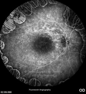
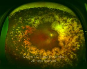
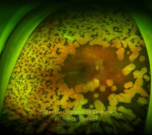
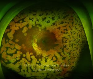
Beginning in the midperiphery there are focal areas of choroidal atrophy with well-circumscribed borders separating normal from abnormal tissue. These lesions progress toward the macula and ora serrata. These areas are initially round to oval and coalesce overtime to form large areas of atrophy, scalloped in appearance, forming a ring or a wreath (hence the term “gyrate”). The borders of the lesions show an extremely sharp demarcation from normal tissue, highlighted by hyperpigmentation. The macula is apparently the most resistant area, with central visual acuity preserved until late in the course of the disease.


