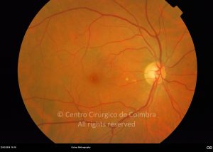
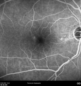
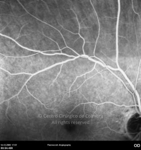
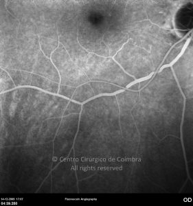
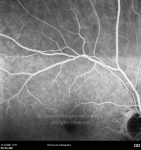
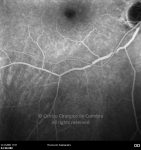
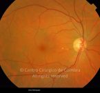
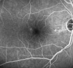
Hypertensive retinopathy is characterized by retinal vascular derangement caused by maintained severe hypertension. High blood pressure causes both focal and generalized constriction of the retinal arterioles due to self-regulation of blood flow.
Chronic elevation of arterial blood pressure and degenerative changes in the arteriolar wall manifest ophthalmoscopically as: retinal arterial attenuation, changes in the arteriolar reflex and arteriovenous crossing changes. Increased vascular permeability can lead to retinal lipid deposits (hard exudates) and flame-shaped hemorrhages. The hemodynamic changes in arterial hypertension can lead to vascular occlusion, focal ischemia and nerve fiber layer infarctions (cotton-wool spots).
Acute severe elevation of blood pressure may lead to acute hypertensive retinopathy (malignant hypertension) and hypertensive choroidopathy. Areas of choroidal infarction can occur, appearing as hyperpigmented lesions surrounded by a halo of hyperpigmentation – Elschnig’s spots. Choroidal infarctions can also assume a linear hiperpigmented configuration along a given meridian – Siegrist’s streaks.
Malignant hypertension is associated with disc swelling and retinal hemorrhages around the disc.


