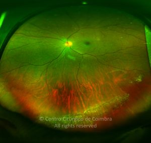
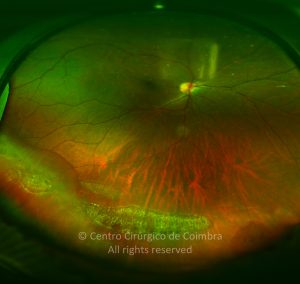
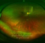
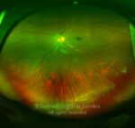
Lattice degeneration is an atrophic disease of the peripheral retina characterized by oval or linear patches of retinal thinning. It is bilateral in one third to one half of affected patients. Condensed vitreous at the margins of the lattice lesions may appear as vitreous opacities and represent regions of increased vitreoretinal adhesion. Retinal tears can occur with posterior vitreous separation pulling on the thinned retina, with an increased risk of retinal detachment. However retinal detachment is a relatively rare complication of lattice degeneration.
Lesions may be isolated or multifocal, variable in dimension, and usually are oriented concentric or slightly oblique to the ora serrata, located within the equatorial region of the fundus, typically inferotemporal.
Patients with lattice degeneration are asymptomatic. The acute onset of floaters, flashes, peripheral field loss, or central vision loss may indicate the presence of a retinal tear or detachment. The vast majority of patients will have lesions that are completely stable or slowly progressive.
It occurs more commonly in but is not limited to myopic eyes.
Snail-track retinal degeneration is a clinical variation of lattice degeneration, occurring mainly in young myopic eyes, characterized by long circumferential areas of retinal thinning with a glistening appearance. Many authors consider it to be early stage of lattice degeneration. The lesions consist of retinal degeneration leading to an atrophy of the tissues with lipid deposits in the internal retinal layers and they have the same complications and clinical protocols.
Differential Diagnosis:
- Retinoschisis
- Atrophic holes
- Chorioretinal scarring
- Congenital hypertrophy of the retinal pigment epithelium
- Retinal holes, tears and detachments


