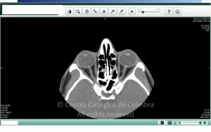
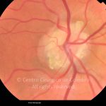
Optic disc drusen are calcified hyaline bodies deposited in the pre-laminar optic nerve. The formation of drusen is related to axonal degeneration of the optic nerve head. Most are congenital, bilateral, and may become visible from the 1st or 2nd decade of life.
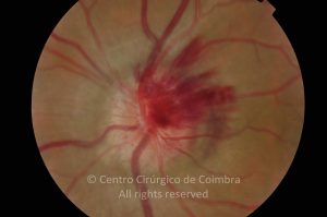
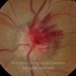
The clinical picture may be characterized by decreased visual acuity and visual field defects. More rarely, drusen can lead to vascular occlusive changes and/or bleeding of the optic disc and retina.
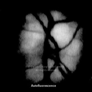
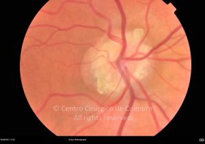
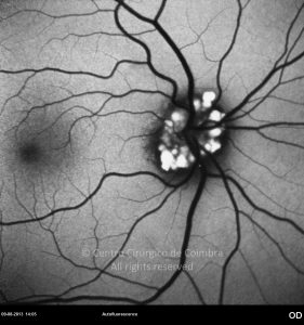
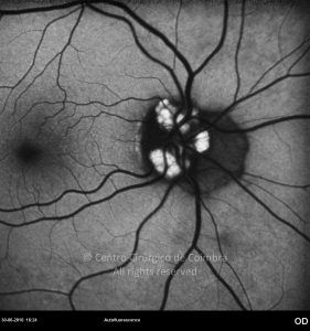
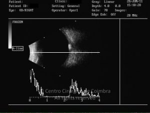
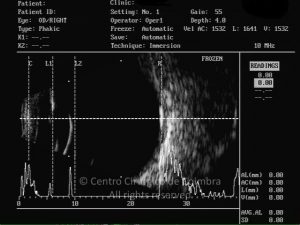
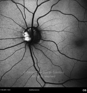
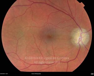
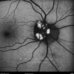
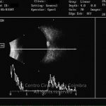
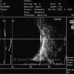
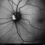
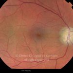
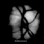
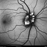
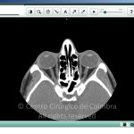
Drusen can be easily identified when they appear as bright yellow hyaline bodies in ophthalmoscopy. When they are inside the nervous tissue of the optic nerve head, they may give a false appearance of optic disc edema (pseudo-papilledema). Superficial drusen are also identified by fluorescein angiography, showing autofluorescence before injection and exhibiting nodular hyperfluorescence after injection. On ultrasonography optic nerve head drusen appear as hyperechogenic with posterior shadowing.


