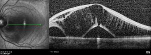
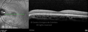
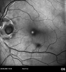
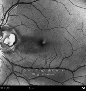
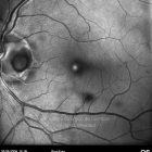
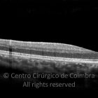
Congenital pit of the optic disc appears as a localized oval or round depression within the nerve head.
Approximately 40% of eyes with congenital optic pit have an associated serous retinal detachment at one time or another, frequently involving the macular region. Chronic retinal detachment can result in lamellar macular holes, cystic retinal degeneration, or retinal pigment epithelial atrophy.
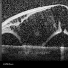
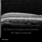
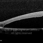
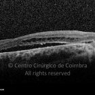
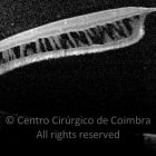
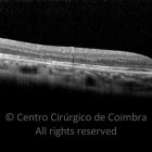
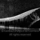
Optical coherence tomography generally reveals a bi-laminar structure, with schisis-like retinal changes overlying a central neurosensory retinal detachment, usually confined to the posterior pole and contiguous with the optic disc.
The origin of the subregional fluid is uncertain: there is speculation that it may arise from the cerebrospinal fluid, the liquefied vitreous, the choroid or the orbit, made easier by vitreous tractions.
In our studies, vitrectomy seems essential in removing these vitreous tractions and we conclude that the anatomical repair may be easier if communication between the pit and the retinal layers is sealed
References
Brown GC, Tasman WS: Congenital Anomalies of the Optic Disc. New York, Grune and Stratton, 1983, pp 95-191.
Travassos A, Travassos AT: Optic Pit: Revisão de Casos Cirúrgicos e Abordagem Terapêutica Inovadora. Revista da Sociedade Portuguesa de Oftalmologia – 2012, Vol35: pp 273-282.


