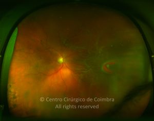
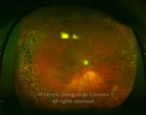
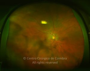
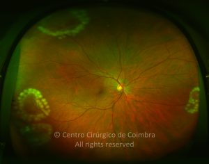
Retinal breaks are full thickness defects of the neurosensorial retina. When significant vitreoretinal traction is present at the break site and a liquefied vitreous is present, a retinal detachment can occur.
Breaks can be caused by atrophy of the inner retinal layers, vitreoretinal traction or trauma. According to their configuration breaks can be divided in: horseshoe tears, giant tears, operculated holes, dialyses, and atrophic retinal holes.
Horseshoe tears, have a U configuration, with the convexity usually pointing to the optic disc. They are caused by vitreous traction, exerted on the flap (frequently the vitreous remains attached to the flap and traction persists). When tears exceed 90 degrees circumferentially they are called giant tears. Tears are most commonly found in the supero-temporal quadrant.
Operculated holes are also caused by vitreoretinal traction, that is usually stronger and released during the hole formation (vitreous remains attached to the operculum and free from attached retina).
Dialyses are circumferential linear breaks occurring either at the anterior or posterior margin of the vitreous base. They are usually found in the infero-temporal retina.
Atrophic holes are caused by atrophy of the inner retinal layers and are not associated with vitreoretinal traction.
The vast majority of retinal breaks do not cause retinal detachment. Therefore treatment must be tailored to specific situations, bearing in mind that the benefits of treatment must be weighted against potential risks.


