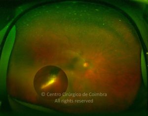
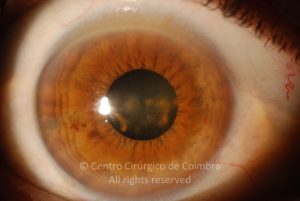
Siderosis bulbi is caused by retention and oxidation of an iron-containing intraocular foreign body. It results from the deposition of iron in the intraocular epithelial structures (lens epithelium, iris and ciliary body epithelium, and sensory retina), where it exerts a toxic effect on cellular enzyme systems, with resultant cell death. Clinical features include cataract, rust-colored anterior subcapsular deposits, iris heterochromia with brownish-discoloration, a dilated and nonreactive pupil, and depressed electroretinogram amplitudes. Other potential sequelae include pigmentary retinopathy, retinal microangiopathy, and open-angle glaucoma.
Siderosis may develop within weeks, but the course is variable depending on the iron content of the foreign body and its location. All ocular structures may be involved in the siderotic process. Functional damage to the retina occurs at a very early stage, before extensive siderosis is apparent, and before stainable iron is detected in retinal tissues. Accumulation of intracellular iron leads to inner retinal and RPE degeneration followed by full-thickness retinal degeneration and secondary gliosis. Vitreous degeneration in association with retinal degeneration causes rhegmatogenous retinal detachment.


