Stargardt disease is the most common juvenile macular dystrophy, generally inherited in an autosomal recessive manner, and characterized by progressive loss of photoreceptors in the macular region leading to loss of central vision.
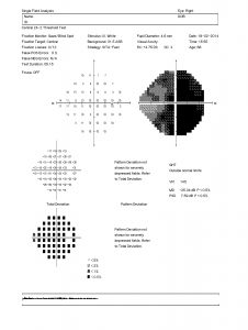
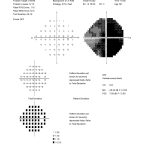
Symptoms begin in the first or second decades of life, with variable progression over time, loss of central vision but with preservation of peripheral vision. Patients complain of dyschromatopsia, usually accompanied by a central scotoma.
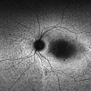
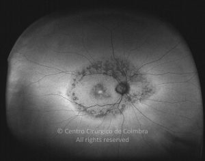
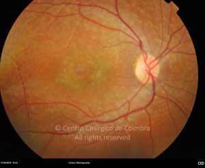
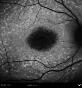
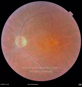
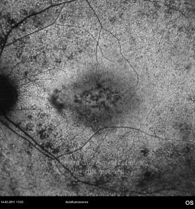
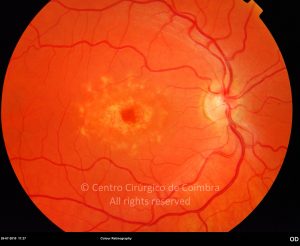
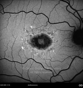
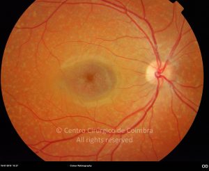
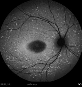
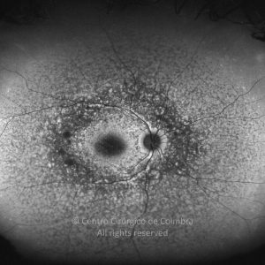
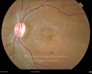
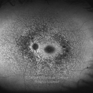
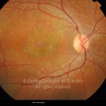
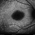
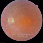
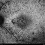
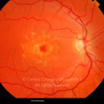
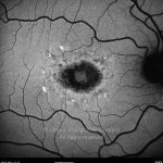
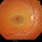
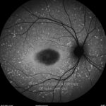
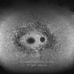
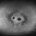
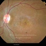
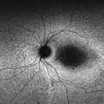
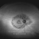
In the initial stages of disease, the fundus is completely normal. Subsequently, changes around the fovea develop, with a granular appearance of the retinal pigment epithelium (RPE). These lesions progress to RPE atrophy and perimacular yellowish fish-tail spots typical of fundus flavimaculatus are observed. These fish-tail lesions are typically hyperautofluorescent (lipofuscin and A2E accumulation) in contrast with atrophic hypoautofluorescent regions, giving sometimes a “bull’s-eye” appearance. The electro-oculography is normal in the early stages and ERG tends to be subnormal.
At present, there is no effective treatment to improve the visual deficit and prevent the progression of this disease. However, ongoing clinical trials with gene therapy and stem-cell technology may represent a great hope in the near future.


