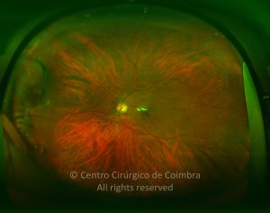
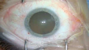
Terson´s syndrome is defined by the occurrence of intraocular hemorrhage secondary to subarachnoid (the most common cause) or subdural hemorrhage. There are several hypotheses to explain this fact. There are several hypotheses to explain this phenomenon. This in turn leads to rupture of the intraocular veins with subsequent hemorrhage.
Hemorrhage can be subretinal, intraretinal, preretinal (subhyaloid) or intravitreal. One particular entity, which can result from the hemorrhage, is a hemorrhagic macular cyst (HMC). HMC is present in up to 40% of patients with Terson syndrome. Two types of HMC have been described Submembranous HMC with blood collected under the ILM and preretinal HMC with blood located between the ILM and the posterior hyaloid. The cyst can have diverse appearances according to its age. Initially it is red, becoming white when blood breakdown has started and transparent when blood breakdown is complete.
Intraocular hemorrhage in the context of intracranial bleeding has prognostic implications. In patients with subarachnoid hemorrhage and Terson syndrome, mortality is two to four times higher than in patients with no intraocular bleeding. Also, the risk of coma in a patient with intracranial bleeding and Terson syndrome doubles when compared with a patient with no intraocular bleeding. Therefore, screening of patients with subarachnoid or subdural hemorrhage under mydriasis should be mandatory.


