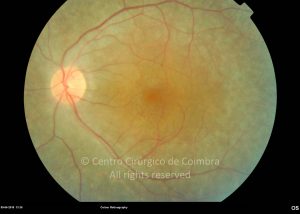
 Fundus photograph of a 32-year-old patient with deafness and poor vision – Usher Syndrome type I. Note the pale disc, extensive areas of RPE atrophy, whitish deposits in the macula periphery and thin vessels.
 Fundus photograph of a 32-year-old patient with deafness and poor vision – Usher Syndrome type I. Note the pale disc, extensive areas of RPE atrophy, whitish deposits in the macula periphery and thin vessels.
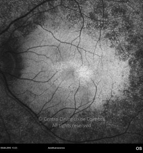
 Autofluorescence image showing a pattern of macular hyperfluorescence, in the same case.

Rua Dr. Manuel Campos Pinheiro, 51 Espadaneira - S. Martinho do Bispo
3045-089 Coimbra, Portugal
(+351) 239 802 700
www.ccci.pt
atlasrleye@ccci.pt
UPS 07/02.00 (Licença de Funcionamento)
Certidão Registo E106499
Imagiologia:LIC-103/2021 (Licença de Funcionamento)