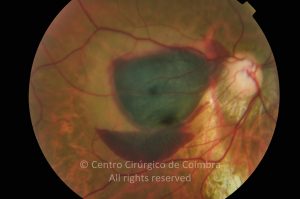
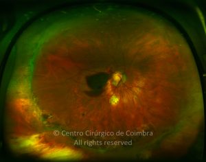
Valsalva retinopathy is characterized by hemorrhage typically located under the internal layer membrane in or near the macular area secondary to a Valsalva maneuver. The sudden increase in intrathoracic pressure that follows the Valsalva maneuver causes a sudden rise in intraocular venous pressure leading to a spontaneously rupture of retinal capillaries. Vitreous hemorrhage and subretinal hemorrhage may be present.
The symptoms range from complains of floating spots with decreased vision to complete loss of central vision. Unilateral manifestations are most common but the findings may be bilateral.
The prognosis for Valsalva retinopathy is generally good and the preretinal hemorrhages usually resolve by themselves in a few weeks to a few months.
Part of the blood turns yellow after several days. Patients with ocular pathologies (acquired retinal vascular abnormalities, congenital retinal vascular disease, moderate and high myopia) are at increased risk for Valsalva retinopathy. Reported causes of a Valsalva maneuver include coughing, weight lifting, vomiting, bungee jumping, aerobic exercise, sexual activity, colonoscopy procedures, fiberoptic gastroenteroscopy, constipation, blowing musical instruments, and compressive injuries.
Differential Diagnosis: Posterior vitreous detachment with hemorrhage; Macronaneurysms.


